Dental X-rays and CBCT scans in brief
Dental X-rays help diagnose problems an examination alone can miss. Examples are decay, gum disease that has reached the bone, and infection at the tip of a root1. A cone beam CT (CBCT) scan shows the teeth and jaws in three dimensions, usually at a higher radiation dose than ordinary dental X-rays2. Each image should be chosen for you, after your history has been taken and you have been examined1.
This page explains the main types of dental imaging and what guidance says about when each is justified. It also covers reported doses, pregnancy and the limits of what images show. Dose figures come from published guidelines and studies, not from our own measurements. On this page "we" means our clinic in Antalya, Turkey. Which imaging equipment is used, and where, is something to ask the clinic; the UK section below lists the questions. The page is part of our technology and laboratory section.
- An X-ray or scan should be chosen for you after your history and an examination, not taken as a routine.
- For decay, gum disease and infection at a root tip, intraoral X-rays give more detail at a lower dose than a panoramic X-ray.
- A CBCT scan is for questions a 2D X-ray cannot answer, such as some implant, wisdom tooth or root canal cases. Guidance does not call for one before every implant.
- CBCT doses are usually higher than 2D dental X-ray doses and vary widely between machines. The smallest suitable area should be scanned.
- Send recent X-rays from your UK dentist, and take home copies of every image in a form your dentist can open.
Why diagnostic imaging may be used
An examination shows the surfaces of the teeth and gums. Much of what shapes treatment lies out of sight: between and inside the teeth, at the root tips and in the bone. Dental imaging is used to look there. It helps diagnose decay, gum disease and infection at the tip of a root1. Three-dimensional scans also have uses in root canal treatment, orthodontics and assessing the jaw before an implant1.
An image is worth taking only if it can change what happens next. The European Commission published guidelines on dental CBCT in 2012 ("the European guidelines" below). They state that a scan should potentially add new information to aid the patient’s management2. Selection Criteria for Dental Radiography (2018) is the UK guidance on choosing dental X-rays ("UK guidance" below). It reminds dentists that X-ray exposure involves a risk to the patient1. Each image is therefore weighed against that risk, as explained under "Clinical justification and optimisation" below.
Images also serve as a record. When an implant crown or bridge is finished, a consensus report recommends an X-ray and probing measurements as a baseline3. Later images are compared with that baseline.
Common dental imaging types
Dental imaging is either two-dimensional (2D), which flattens the teeth and jaws into one picture, or three-dimensional (3D).
- Intraoral X-rays are taken with a small sensor or film held inside the mouth. A bitewing shows the crowns of the back teeth and the bone between them. A periapical X-ray shows one or a few whole teeth, down to the root tips.
- Extraoral 2D X-rays are taken with the sensor outside the mouth. A panoramic X-ray (also called an OPG) shows both jaws and all the teeth in one wide image. A cephalometric X-ray is a side view of the skull, used in orthodontics.
- Cone beam CT (CBCT) produces high-resolution, three-dimensional images of teeth and jaws1. The scanned area, called the field of view, can be limited to the teeth and jaws or extend to the face2.
- Medical CT and MRI are hospital scans. When they are used instead of CBCT is explained under limitations below.
An intraoral scan, used to make crowns, bridges or aligners, is a different thing. It records the surface of the teeth and gums, and is covered on our page on digital scanning.
| What it shows | Typical uses | |
|---|---|---|
| Bitewing (intraoral) | The crowns of the back teeth and the bone level between them | Decay between the back teeth; bone level in gum disease |
| Periapical (intraoral) | One or a few whole teeth, including the root tips | Root canal treatment, infection at a root tip, checking an implant |
| Panoramic (OPG) | Both jaws and all the teeth in one image, with less fine detail | An overview of the whole mouth |
| Cephalometric | A side view of the skull and jaws | Orthodontic assessment |
| CBCT | A 3D volume of part of the jaws, or of the face | Selected implant, root canal, wisdom tooth and injury cases |
| Medical CT (hospital) | Bone and soft tissues | When soft tissues need assessing |
| MRI (hospital) | Soft tissues | When soft tissues need assessing |
When two-dimensional imaging may be sufficient
For most everyday questions, a 2D X-ray is the first choice. UK guidance finds intraoral X-rays better than panoramic X-rays for diagnosing decay, gum disease and infection at the root tip1. For these it calls intraoral X-rays the optimal examination, because of better detail and lower radiation doses1.
The European guidelines date from 2012 and recommended their own review within five years. Read them with that age in mind. They say a scan should be used only when the question cannot be answered adequately by lower dose conventional (traditional) radiography2. They list situations where CBCT is not the standard method2:
- finding decay;
- checking bone support in gum disease, as a routine;
- diagnosing infection at the tip of a root, or showing the shape of the root canals, as a standard method. The guidelines note that an operating microscope may reveal canal anatomy without radiation;
- assessing every wisdom tooth before removal, as a routine;
- large-volume scans for orthodontic diagnosis, as a routine.
After an implant has been placed, recommendations say 2D intraoral X-rays are still the main tool4 when it needs checking. So your own dentist in the UK can usually follow an implant with standard X-rays.
When CBCT may be considered
A CBCT scan may be justified when a 2D X-ray leaves open a question that matters for treatment. The European guidelines name selected cases2, each where ordinary X-rays do not give enough information:
- a suspected root fracture after an injury to a tooth;
- root canals whose shape stays unclear, most often in back teeth with several roots;
- some bone defects around teeth affected by gum disease;
- teeth that have not come through, before surgery;
- a lower wisdom tooth that is to be removed, where X-rays suggest its roots are in close contact with the nerve canal.
Our pages on wisdom tooth removal and root canal treatment explain those treatments.
Before implants. The European guidelines accept CBCT before an implant as an alternative to other cross-sectional (3D) methods. The condition is that the CBCT dose is shown to be lower. They give that recommendation their lowest evidence grade (D)2. They add that the first question is whether cross-sectional imaging is needed at all. Recommendations from 2018, based on a narrative review, say a scan could be justified for diagnosis and planning before surgery4. Neither source says that every implant needs a CBCT scan. Whether you need one depends on the site, the bone and what the 2D images show. See dental implants, and digital treatment planning for how a scan is used in planning software.
Clinical justification and optimisation
Two principles apply to every dental X-ray and scan.
Justification. The expected benefit has to outweigh the risk, for you as an individual. UK guidance calls this both an ethical and a legal requirement1. It describes X-rays taken by a general rule, rather than prescribed for the individual, as unacceptable1. It defines a routine X-ray as one taken whether or not there are signs or symptoms. Routine X-rays of this kind cannot be justified1. Check-up X-rays chosen for you are different. In checking for decay, your assessed risk is central to deciding when further X-rays are taken1. The interval should be specific to each patient1. For CBCT, the European guidelines add that each scan must be justified for each patient2. It should not be repeated ‘routinely’2 without a new assessment of benefit and risk.
Optimisation. Once an image is justified, the dose should be as low as still gives a reliable answer. For CBCT, that means the smallest scanned volume compatible with the clinical situation2. It also means the resolution compatible with adequate diagnosis and the lowest achievable dose2. The 2018 implant recommendations describe the aim as a dose as low as diagnostically acceptable4. Equipment matters too. The European guidelines found that some dental CBCT equipment is associated with effective doses that are not as low as reasonably achievable2.
You can ask why an image is needed, what question it should answer, and whether an image you already have would do.
Radiation exposure and protection
Dental X-rays and CBCT scans use ionising radiation. According to UK guidance, the risk is mainly that of causing cancer1. It is highest for the young and lowest for the elderly1.
Doses are compared as "effective dose", in microsieverts (µSv). Effective doses are calculated for a reference patient, with many uncertainties1. They are not a measurement of your own dose. The table shows ranges from the European guidelines’ 2012 tables, based on measurements on models (phantoms)2. The guidelines warn that such figures become dated very quickly2.
| Effective dose in µSv (median in brackets where reported) | |
|---|---|
| One intraoral X-ray | Under 1.5 (with a rectangular beam; almost five times higher with a round one) |
| Panoramic X-ray | 2.7 to 24.3 |
| Cephalometric X-ray | Under 6 |
| CBCT of the teeth and jaws (small or medium field) | 11 to 674 (61) |
| CBCT of the face (large field) | 30 to 1,073 (87) |
| Medical CT of the jaws | 280 to 1,410 |
Later sources show the same wide spread. A 2015 meta-analysis also used model measurements. In adults, it found doses of 5 to 652 µSv for small fields, 9 to 560 for medium and 46 to 1,073 for large fields5. Its authors concluded that such wide ranges make CBCT doses hard to generalise. A 2018 review reports CBCT doses equal to between 2 and 200 panoramic X-rays, even for similar purposes4. So there is no single "CBCT dose". It depends on the machine, its settings and the size of the area scanned. In general, CBCT doses are higher than those of intraoral and panoramic X-rays, and lower than medical CT2.
Protection. The main protections are the ones described above: no image without a reason, no unnecessary repeats, and the smallest suitable scan. Advice on shields has changed over time and still differs. UK guidance from 2018 sees no need for the routine use of lead aprons1 in CBCT, but finds some evidence for a thyroid shield. A later UK joint report on patient shielding (2020) does not recommend shielding for dental radiography for the majority of imaging situations6. For CBCT it makes an exception for large-field scans, where there may be some benefit. It warns that a thyroid collar may lead to artefacts in the images6. A shield can also interfere with the imaging, leading to a repeat test6. A 2023 recommendation from the American Academy of Oral and Maxillofacial Radiology advises that thyroid shielding not be used7 for dental X-rays or scans. Ask what the dentist uses and why.
Pregnancy and medical-history considerations
Pregnancy. Tell the dentist before any X-ray if you are or might be pregnant. According to UK guidance, dental X-rays, including panoramic and CBCT scans, do not need to wait until after the birth1. It advises abdominal protection only for one rarely used view, the vertex occlusal X-ray, which points the beam at the abdomen1. You may also choose to postpone an X-ray that is not urgent1. The 2020 UK joint report is more cautious about CBCT. It notes that the reasons for a CBCT scan are limited. For a pregnant patient, it may be more appropriate to postpone imaging until after pregnancy6. A 2024 systematic review concluded that dental imaging in pregnancy should not be restricted if clinically indicated8. It also noted that few studies have looked at this.
Guidance on shielding in pregnancy differs. The 2023 US radiology recommendation advises discontinuing fetal shielding7 for dental imaging. A 2013 US obstetric committee opinion recommends shielding of the abdomen and thyroid9. Whether or not an X-ray is taken, UK guidance recommends recording your agreement to that decision1 in your notes.
Your medical and dental history. A history and an examination come before any scan. The European guidelines say a CBCT scan must not be carried out2 without them. Tell the dentist about X-rays and scans you have had recently, and bring them. UK guidance advises dentists to seek originals or copies of radiographs taken elsewhere1 when they are relevant. It also asks them to avoid images that duplicate those taken previously1. Mention any metal crowns, posts or fillings too: metal can blur a CBCT scan, as explained below.
Image interpretation and limitations
An image is only as useful as its reading.
- The whole scan is read. The European guidelines call for a thorough clinical evaluation (‘radiological report’) of the entire image dataset2, not only of the area being treated. Large scans often include the base of the skull. UK guidance says these need to be viewed by a suitably qualified radiologist1 to rule out unexpected findings.
- Who reports. For scans of the teeth and jaws, the European guidelines recommend a report by a specially trained dento-maxillofacial radiologist2. Where that is impracticable, they accept an adequately trained general dental practitioner2. Ask who will report your scan, and whether you will receive the written report.
- Detail. CBCT gives the advantage of three dimensions at the expense of a loss of resolution1, compared with intraoral X-rays.
- Metal. Metal crowns, posts and fillings can cause streaks on CBCT. These artefacts1 can look like decay or hide it, and can profoundly reduce image quality1.
- Soft tissues. CBCT is designed for teeth and bone. Where soft tissues need assessing, the guidelines advise conventional medical CT or MR, rather than CBCT2.
- Measurements. Distances measured on CBCT are comparable in accuracy with CT1. But bone density values read from CBCT are not reproducible1. The European guidelines add that accuracy in patients may be lower than in laboratory studies, because of small movements during the scan. They advise clinical judgement and a margin of safety2 when planning implants close to important structures.
- Planning. A scan shows where the nerve canal and the sinus lie, so treatment can be planned around them. It does not remove the risks of surgery.
An image never replaces an examination. A normal-looking X-ray does not rule out every problem: the dentist reads it together with your symptoms, the examination and any tests. Any opinion given on X-rays or scans you send is preliminary until you have been examined.
Records, consent and referrals
Records. Every image, and the report on it, is part of your dental record. In Turkey, the health tourism regulation entitles international patients to an itemised bill. On request, they also get free copies of the records of the materials used, the tests and the imaging10. Patients may also examine their file and take a copy11. A CBCT scan is a set of data files, not a single picture. UK guidance notes that most CBCT makers provide an export function with a simple imaging program1, so the data can be viewed elsewhere. Ask in which format you will receive your images.
Consent. You can ask why an image is needed, what it should show and what would happen if you said no. In Turkey, patients are entitled to information on other options, with their benefits and risks, and the consequences of refusing11. If a decision to take an X-ray, or not, could be disputed, UK guidance says a record should be made of that discussion and decision1.
Referrals. Sometimes one dentist asks another, or a hospital, to take an image. UK guidance says the request should carry adequate clinical information. A note such as “please take a panoramic radiograph for diagnosis” is not enough1. For CBCT, the referring dentist must supply the results of a history and examination2. If a scan raises a question beyond the teeth and jaws, or soft tissues need assessing, you may be referred for hospital imaging.
If you live in the UK: questions and records
Before you travel
- Send X-rays or scans from your UK dentist, with the date each was taken. If they answer the treating dentist’s question, a repeat may be avoided. An opinion based on them is preliminary.
- Ask the clinic in writing which images it expects to take, and why.
- Ask whether images are taken at the clinic or at an outside imaging centre.
- Ask who will report a CBCT scan.
What to bring home
- Copies of every X-ray and scan, with their dates, in a form your UK dentist can open. For a CBCT scan, ask for the data files with a viewer, and the report.
- The written treatment record that goes with them.
Afterwards. Later problems are usually first seen by your own UK dentist, who will need these images. If you do not have one, it helps to find one before you travel. The General Dental Council (GDC) suggests speaking to your own dentist12 before you consider treatment abroad. The NHS website for England lists exchanging medical records and arranging aftercare back home13 among the things to consider. It also says the NHS is not liable for negligence or failure of treatment13 abroad. It adds that most travel insurance policies will not cover you for planned treatment abroad13. That is guidance for England. Scotland, Wales and Northern Ireland run their own health services, with their own rules, which this page does not cover; if you live there, ask your dentist.
Your choice. You can ask your UK dentist whether an X-ray you need could be taken in the UK before you go. You can say no to an image, or to treatment.
The questions the GDC and the NHS suggest you ask are on our page on dental treatment in Turkey.
Frequently Asked Questions
Do I need a CBCT scan before a dental implant?
Not always. Guidance says a 3D scan can be justified for planning an implant. The first question is whether 2D X-rays already answer what the dentist needs to know. The decision is made for you after an examination.
Is a CBCT scan the same as an intraoral scan?
No. A CBCT scan uses X-rays to show the teeth, roots and jawbone in three dimensions. An intraoral scan records only the surface of the teeth and gums, for making crowns, bridges or aligners.
Can I have dental X-rays while pregnant?
Yes, if they are needed. UK guidance says dental X-rays, including panoramic and CBCT scans, need not wait until after the birth. A 2020 UK report suggests a CBCT scan may be better postponed. Tell the dentist if you are or might be pregnant.
Can I send X-rays my UK dentist has already taken?
Yes. Send X-rays or scans with the date each was taken. The treating dentist decides whether they are recent enough to answer the question; if so, a repeat may be avoided. Any opinion based on them stays preliminary until you are examined.
Will I get copies of my X-rays and scans?
Ask for copies of every image before you leave, in a form your UK dentist can open. For a CBCT scan, ask for the data files with a viewer and the written report. In Turkey you can request copies of your imaging records.
Who reads my CBCT scan?
The whole scan should be reported, not only the area being treated. Ask who will report yours, a dental radiologist or a trained dentist, and whether you will receive the written report.
Should I wear a lead apron or a thyroid collar?
Guidance differs. UK guidance from 2018 sees no need for routine lead aprons in CBCT but some evidence for thyroid shields. A 2020 UK report and a US recommendation advise against shielding in most dental imaging. Ask the dentist what they use and why.
How much radiation does a dental X-ray give?
It depends on the type. A single intraoral X-ray usually gives the lowest dose and a panoramic X-ray more, though several X-rays add up. A CBCT scan usually gives more again, with a wide range between machines. The dose table on this page gives published ranges.
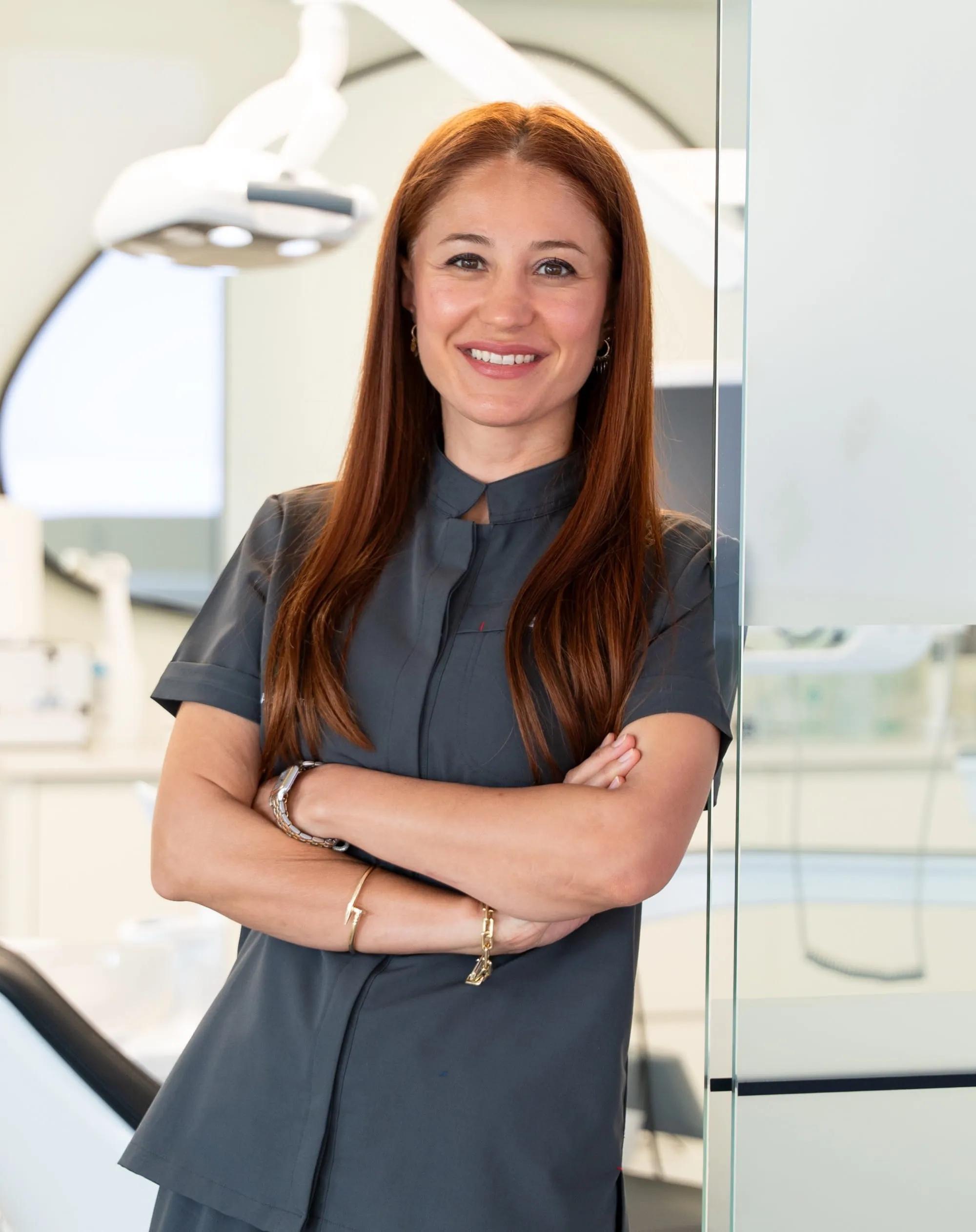
Dt. Dilek AKSU GÜLER
Dentist · Co-founder
Dt. Dilek AKSU GÜLER qualified from the Faculty of Dentistry at Süleyman Demirel University, Turkey, in 2005. She is the founding dentist of our clinic in Lara, Antalya. She works in aesthetic restorations (veneers and crowns), digital smile design and implant-supported prostheses, and speaks Turkish, English and German.
Sources
- Selection Criteria for Dental Radiography, 3rd edition. Faculty of General Dental Practice (UK), now College of General Dentistry, updated 2018, eds Horner K, Eaton KA. 2018.↩cgdent.uk
- Radiation Protection No 172. Cone beam CT for dental and maxillofacial radiology: evidence-based guidelines. European Commission, Directorate-General for Energy (SEDENTEXCT project, Euratom FP7), Luxembourg 2012. 2012.↩op.europa.eu
- Peri-implant diseases and conditions: consensus report of workgroup 4 of the 2017 World Workshop on the Classification of Periodontal and Peri-Implant Diseases and Conditions. Journal of Clinical Periodontology 2018;45 Suppl 20:S286-S291. 2018.↩doi.org
- Cone beam computed tomography in implant dentistry: recommendations for clinical use. BMC Oral Health 2018;18(1):88 (Jacobs R, Salmon B, Codari M, Hassan B, Bornstein MM). 2018.↩doi.org
- Effective dose of dental CBCT: a meta analysis of published data and additional data for nine CBCT units. Dentomaxillofac Radiol 2015;44(1):20140197 (Ludlow JB et al.); erratum DMFR 2015;44(7):20159003. 2015.↩doi.org
- Guidance on using shielding on patients for diagnostic radiology applications. British Institute of Radiology, joint report of BIR, IPEM, Public Health England, RCR, SCoR and SRP (London, March 2020). 2020.↩bir.org.uk
- Patient shielding during dentomaxillofacial radiography: recommendations from the American Academy of Oral and Maxillofacial Radiology. Journal of the American Dental Association 2023;154(9):826-835.e2. 2023.↩doi.org
- Impact of dental imaging on pregnant women and recommendations for fetal radiation safety: a systematic review. Imaging Science in Dentistry 2024;54(1):1-11. 2024.↩doi.org
- Oral health care during pregnancy and through the lifespan (Committee Opinion No. 569). American College of Obstetricians and Gynecologists, Obstet Gynecol 2013;122:417-22, reaffirmed 2025. 2013.↩acog.org
- Uluslararasi Saglik Turizmi ve Turistin Sagligi Hakkinda Yonetmelik. T.C. Saglik Bakanligi, Resmi Gazete 26/4/2025, No 32882. 2025.↩resmigazete.gov.tr
- Hasta Hakları Yönetmeliği (consolidated text). T.C. Sağlık Bakanlığı; Resmî Gazete 1/8/1998 No 23420, amended RG 8/5/2014-28994, 23/12/2016-29927, 16/1/2019-30657. 1998.↩mevzuat.gov.tr
- Going abroad for dental treatment (patient information). General Dental Council, accessed 18 September 2026. 2026.↩gdc-uk.org
- Going abroad for medical treatment. NHS (England), accessed 21 September 2026.↩nhs.uk